Simple renal cyst
In 1 minute
A simple renal cyst, which is often what people mean by a "regular renal cyst," is the classic benign simple-fluid cyst pattern rather than just any cystic space in the kidney. On ultrasound it is typically round or oval, thin-walled, anechoic, and shows posterior acoustic enhancement; on CT or MRI it behaves like simple fluid and does not show enhancing septa, nodules, or solid parts.682
The main radiology question is not just "is there a cyst?" but "is it truly simple, or is something making it complex?" Once there is hemorrhagic or proteinaceous content, septa, calcification, wall irregularity, enhancement, parapelvic-location confusion, or a broader multiple-cyst context, the simple cyst story weakens and the workup changes toward contrast CT or MRI characterization.246
Fast engineer rule: preserve modality, phase, multiplicity, and the exact words simple versus complex. If a lesion is truly simple, it is benign and usually needs no follow-up. Slow down when there are internal echoes or debris, septa, enhancement, parapelvic collecting-system uncertainty, symptoms, or many bilateral cysts, because those findings push the workflow out of the routine simple-cyst lane.125
Quick diagnostic logic
| Situation | What radiologists look for | Why it matters |
|---|---|---|
| Classic simple cyst on ultrasound68 | Round or oval shape, thin wall, anechoic center, no septa, and posterior acoustic enhancement | This is the clean ultrasound pattern that supports a true simple cyst |
| Simple cyst on CT or MRI21 | Water-like attenuation or signal, hairline-thin wall, no calcification, no septa, no solid component, and no enhancement | This is the Bosniak I / benign simple-cyst lane, where follow-up is usually not recommended |
| Lesion is not simple42 | Septa, wall thickening, internal echoes or debris, calcification, mural nodules, or enhancement | These features move the lesion toward hemorrhagic, proteinaceous, or complex cystic mass logic rather than a regular simple cyst |
| Parapelvic cyst versus hydronephrosis6 | Whether the cystic spaces are discrete and rounded or whether they branch through the collecting system like hydronephrosis | Hydronephrosis follows collecting-system anatomy; a parapelvic cyst does not, and mixing them up can change the whole workflow |
| Multiple bilateral cysts raise a broader cystic-disease question265 | Kidney size, family history, CKD or dialysis context, and extra-renal cysts | Many bilateral cysts are not the same thing as one incidental simple cyst and may suggest ADPKD or ACKD |
| Symptomatic cyst or management trigger27 | Pain, infection, hemorrhage, obstruction, hematuria, and whether size or location explains symptoms | Most simple cysts need nothing, but symptomatic cysts can move toward aspiration with sclerotherapy or laparoscopic deroofing |
Example figures
These are lesion-specific simple renal cyst ultrasound examples from open-access PMC-hosted articles. Both images show the key teaching point for this page: a true simple cyst is anechoic and produces posterior acoustic enhancement.86
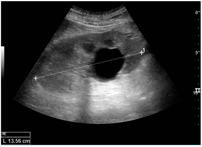
Figure 1. Simple renal cyst with posterior acoustic enhancement from an open-access ultrasound pictorial review. This is the clean adult-kidney ultrasound pattern radiologists mean when they call a cyst truly simple. Source:
Sorensen et al., Figure 5
, CC BY 4.0.8
If you want the most practical pitfall comparison for this page, see
Figure 8 from My patient has abdominal and flank pain: Identifying renal causes
. It shows a simple parapelvic cyst that does not communicate with the renal pelvis, which is exactly the visual clue that helps separate parapelvic cyst from hydronephrosis on ultrasound.9
For the comparison case, see
Figure 16 from Ultrasonography of the Kidney: A Pictorial Review
. It shows hydronephrosis as an interconnected anechoic pelvis-and-calyces pattern with cortical atrophy, which is the opposite of a discrete parapelvic cyst that does not branch through the collecting system.8
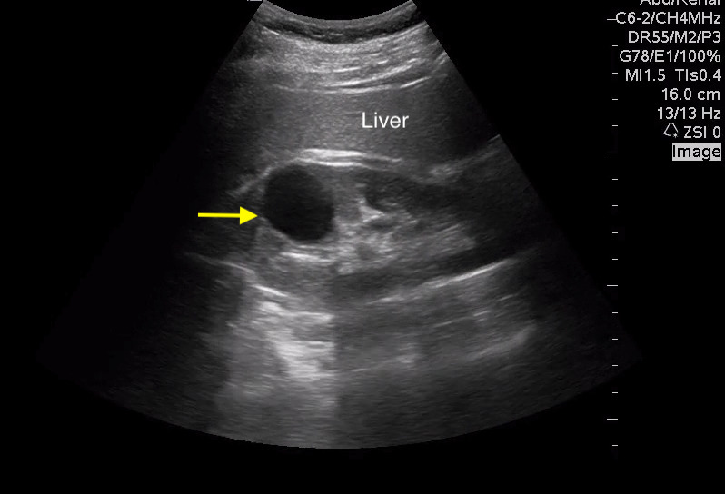
Figure 2. Point-of-care ultrasound example of a simple renal cyst in the right kidney. This figure is useful because it reinforces the same simple-cyst pattern in a practical bedside ultrasound setting: oval fluid content, no internal complexity, and posterior enhancement. Source:
Koratala et al., Figure 7
, CC BY 4.0.6
Common confusions
- Hemorrhagic or proteinaceous cyst: these may still be benign, but once the cyst contains blood or protein it stops looking like simple fluid. That is why internal echoes on ultrasound, high attenuation on CT, or T1-bright content on MRI push the reader away from the ordinary simple-cyst explanation.4 See also hemorrhagic renal cyst.
- Complex cystic mass / Bosniak > I: septa, calcification, wall thickening, measurable enhancement, or nodularity mean the lesion is no longer a regular simple cyst. At that point the question becomes Bosniak category and malignancy risk, not "simple cyst or not?"12
- Cystic RCC: a cystic renal cancer can still look partly fluid-filled, but enhancing septa, wall thickening, or mural nodules are exactly the features that keep radiologists from calling it simple.43
- Parapelvic cyst versus hydronephrosis: parapelvic cysts can look like a dilated collecting system, especially on limited ultrasound. Hydronephrosis tends to branch through the renal sinus and calyces, while cysts are discrete rounded spaces that do not follow the collecting-system pattern.6
- ADPKD versus multiple incidental simple cysts: multiple bilateral cysts, enlarged kidneys, family history, and liver cysts should shift attention away from the "one regular cyst" mindset and toward inherited cystic kidney disease.26
- ACKD versus incidental simple cysts: in advanced CKD or dialysis patients, multiple cysts in smaller chronically diseased kidneys suggest acquired cystic kidney disease rather than just age-related simple cysts.65
Engineer-first takeaway
If you only remember one workflow rule, remember this: do not collapse all renal cysts into one label. The important handoff is whether the lesion was reported as simple, complex, hemorrhagic/proteinaceous, parapelvic, multiple bilateral, or symptomatic. That wording tells you whether radiologists thought the lesion stayed in the benign incidental lane or had already moved into a more complex renal-cyst workflow.26
Ultrasound is good at recognizing a classic simple cyst, but it is not the same thing as contrast CT or MRI characterization of a lesion that is no longer obviously simple. For engineering, NLP, and structured-data work, it helps to preserve nearby distinctions such as:
- simple renal cyst
- complex renal cyst
- hemorrhagic cyst
- Bosniak I
- Bosniak II or IIF
- parapelvic cyst
- multiple bilateral renal cysts
- symptomatic renal cyst
Compared with renal mass pages such as RCC, the true simple-cyst literature is much more about classification, differential, and symptomatic treatment than about dedicated public datasets or AI benchmarks. That gap is an EnginRad inference from the source landscape on this page rather than a direct claim from one paper.27
The dataset reality is even narrower than the imaging literature. We did not find a credible public benchmark specifically labeled for true simple renal cyst. The closest partial-fit resource is KiTS23, which includes kidney, tumor, and cyst segmentation targets on contrast-enhanced CT, but it is still an oncology- and nephrectomy-oriented segmentation challenge rather than a benign incidental simple-cyst dataset.10 A broader fallback is ULS23, which includes kidney lesions inside a universal CT lesion segmentation benchmark, but it does not provide Bosniak-style semantics or a simple-cyst-versus-not-simple-cyst label space.11
That means the main technical gap here is not basic recognition of what a simple cyst looks like. The real gap is the absence of a public benchmark that jointly preserves simple cyst, hemorrhagic or proteinaceous cyst, parapelvic cyst, hydronephrosis mimic, multiple bilateral cysts, and ADPKD or ACKD pattern as separate labels.1011
Related research map
Adult workflow cloud
Current practical papers for incidental simple-cyst language, referral, and what changes once symptoms matter.
Boundary and classification cloud
Papers that define where a lesion stays truly simple versus when broader cystic-mass logic takes over.
Ultrasound and pitfall cloud
Visual and bedside sources for recognizing a true simple cyst and spotting the common look-alikes.
Benchmark gap cloud
Closest public data is partial-fit only. There is still no clean public benchmark for true simple renal cyst labels.
Fresh pipeline candidates
Auto-generated from the latest paper-engine review. These papers are visible here before they are manually curated into the main map.
2023
KiTS23 partial-fit benchmark
This is the closest public kidney dataset that explicitly includes cyst targets, but it is still a nephrectomy and oncology segmentation challenge rather than a benign incidental simple-cyst benchmark.
2024
ULS23 broader lesion fallback
A broader universal CT lesion benchmark that includes kidney lesions, but only as part of a much larger lesion-segmentation task.
Clinical and pathology background
A simple renal cyst is an epithelial-lined, fluid-filled renal lesion that represents the benign end of the renal-cyst spectrum. Current review literature describes kidney cyst formation as a process involving focal tubular epithelial proliferation, dilation, and outpouching; in the simple-cyst setting, the result stays a clean fluid lesion rather than a hemorrhagic or complex mass.2 Because the contents behave like simple fluid, ultrasound shows an anechoic lesion with posterior acoustic enhancement, and CT or MRI show a lesion without the enhancing internal features that would move it into a more suspicious category.81
Most simple renal cysts in adults are incidental, benign, and asymptomatic. That is why the routine story is so calm: once the cyst is truly simple, the main job is usually to recognize it correctly and stop there rather than escalating the workup.25 The clinical background changes when symptoms appear, which can happen because of pain, hemorrhage, infection, hematuria, stretching of the capsule, or obstruction, and is influenced by both cyst size and location.76
Once a simple cyst becomes clearly symptomatic, treatment options such as aspiration with sclerotherapy and laparoscopic deroofing or decortication enter the conversation. The current meta-analysis suggests that aspiration with sclerotherapy has higher symptomatic and radiologic failure, while also having fewer complications, shorter stay, and lower cost than laparoscopic deroofing.7 That means the management story is a tradeoff rather than a single always-best answer.
The broader cystic-disease differential matters because not every patient with multiple cysts simply has "a lot of simple cysts." ADPKD usually points toward enlarged kidneys, family history, and often extra-renal cysts such as in the liver. ACKD is more tied to advanced CKD or dialysis and tends to sit in smaller chronically diseased kidneys.265 That is why multiplicity and kidney background are part of the simple-cyst workflow, not just side notes.
Scope and caution
This page is educational and intentionally simplified for technical readers. It does not replace formal radiology interpretation, urology or nephrology evaluation, local policy, or patient-specific medical advice.
References
- Bosniak Classification of Cystic Renal Masses, Version 2019: https://pmc.ncbi.nlm.nih.gov/articles/PMC6677285/
- A clinician's guide to the diagnosis and management of kidney cysts: https://pmc.ncbi.nlm.nih.gov/articles/PMC12515459/
- Management of the Incidental Renal Mass on CT: A White Paper of the ACR Incidental Findings Committee: https://geiselmed.dartmouth.edu/radiology/wp-content/uploads/sites/47/2019/04/ACR_Renal2017.pdf
- CT and MR imaging of cystic renal lesions: https://pmc.ncbi.nlm.nih.gov/articles/PMC6942066/
- How simple are 'simple renal cysts'?: https://pmc.ncbi.nlm.nih.gov/articles/PMC4158337/
- Bedside Assessment of the Kidneys and Bladder Using Point of Care Ultrasound: https://pmc.ncbi.nlm.nih.gov/articles/PMC9994308/
- Comparison of aspiration with sclerotherapy and laparoscopic deroofing for the treatment of symptomatic simple renal cysts: a systematic review and meta-analysis: https://pmc.ncbi.nlm.nih.gov/articles/PMC8500865/
- Ultrasonography of the Kidney: A Pictorial Review: https://pmc.ncbi.nlm.nih.gov/articles/PMC4808817/
- My patient has abdominal and flank pain: Identifying renal causes: https://pmc.ncbi.nlm.nih.gov/articles/PMC4760602/
- 2023 Kidney and Kidney Tumor Segmentation Challenge: https://doi.org/10.5281/zenodo.7840134
- The ULS23 Challenge: a Baseline Model and Benchmark Dataset for 3D Universal Lesion Segmentation in Computed Tomography: https://arxiv.org/html/2406.05231v1